A Breakthrough, First Cryo-EM Structure from India
Recently, scientists from NCCS, Pune have studied a protein with the help of Cryo-EM to gain its structural and functional insights. It is considered to be the first Cryo-EM based structure determination study, carried entirely in India. The study has been published in Scientific Reports.
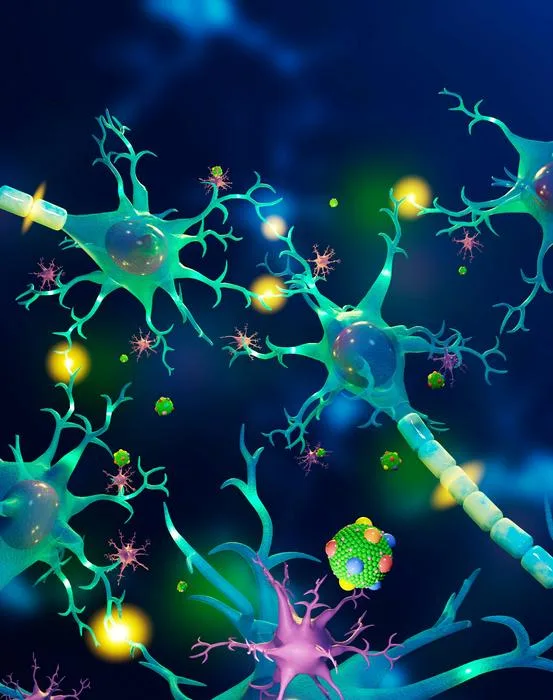
Most of the neurological disorders involve dysfunctioning of receptors which plays a crucial role in the transmission of the synaptic signals. One such important receptor is GluK3 Kainate located at the pre and post synapse. The scientist tried to understand the gating properties unique to the receptor which helps in the regulation of synaptic transmission across the neurons.
Here we share the interview the Dr. Janesh Kumar (Scientist) addressing important insight about the study.
How did you come up with this hypothesis & what got you interested?
Dr. Janesh – “GluK3 receptors belong to the family of glutamate receptor ion channels and play critical roles in maintaining the balance between excitatory and inhibitory neurotransmission, which is essential for normal brain functions. However, our understanding of how they work and could be regulated was limited due to a lack of detailed 3D view for these receptors. Hence, we decided to work on it to fill this gap. I got interested in the area of membrane protein biology in general and ion channels in particular in 2003 when I heard Rob Mackinnon’s Nobel lecture. The interest got reinforced during my postdoctoral stint at NIH, Bethesda with Dr. Mark Mayer.”
Why is your research important? What are the possible real-world applications?
Dr. Janesh -“Typical functions of the brain depend on the ability of nerve cells to transmit electrical signals. Glutamate receptor ion channels are molecular machines that are fundamentally involved in this electrical transmission in the brain. Dysfunction of these ion channels is implicated in a remarkable range of diseases of the nervous system such as Alzheimer’s, Parkinson’s, epilepsy, schizophrenia, etc. So it’s critical to understand how these receptors operate and how their functions are regulated. Towards this goal, we trapped the receptor in two different states in the gating cycle and determined their structure via electron microscopy. The comparison and analysis of the structures revealed the various movements in the receptor molecule responsible for its functions. Using these structures as a guide, we show that the sugar chains that are present on the receptor surface mediate interactions between various regions of the receptor and tune receptor functions. Our results provide the first 3D view of GluK3 receptors and show that sugars present on the receptor surface modulate their functions.”
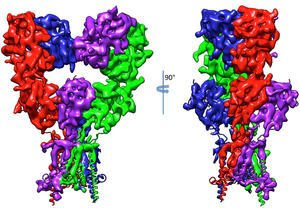
Credit – Dr. Janesh, NCCS, Pune
“This detailed view gives critical clues to developing drugs for combating several neurological diseases and conditions. It is invaluable as efforts are ongoing around the world to develop drugs that might work on specific receptor subunits that may help fight or cure some of the neurological diseases and conditions.”
What kind of response have you gotten to your research /findings?
Dr. Janesh -“We have received a lot of appreciation for this work from all corners. This is primarily because of the crucial roles these receptors play in the brain but also because working with membrane proteins is considered very challenging. This is due to their weak expression combined with their instability when extracted out of cell membranes. Further, our work reports the first structure of a eukaryotic membrane protein from India. It’s also probably the first cryo-EM structure reported from India (where the entire work is carried out in India)”.
What are the challenges faced during the discovery?
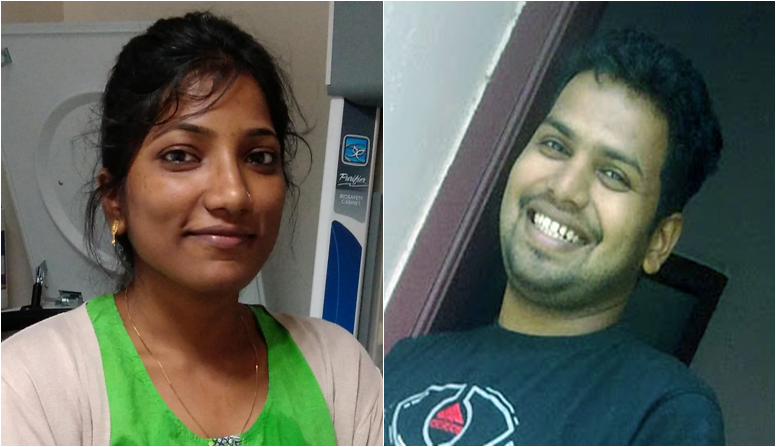
Dr. Janesh -“There were multiple hurdles on the way. Beginning with setting up a facility for membrane protein expression and purification from eukaryotic cells to set up sophisticated instrumentation for doing electrophysiology experiments at NCCS Pune. Talented and hardworking lead authors on the paper who did the entire work reported in this paper, Ms. Jyoti Kumari and Dr.Rajesh Vinnakota took up this challenge and successfully established the respective facilities in the lab. We have now helped many other labs in India in establishing similar methods for protein expression and purification. This could not have been possible without their efforts and the funding support from India Alliance and DBT, New Delhi”.
“However, our biggest challenge has been getting enough time on high-end cryo-electron microscopes for performing these experiments. Let me elaborate a bit on this. Before a large dataset could be collected for high-resolution structure determination, one must identify/screen suitable conditions for EM experiments. For single-particle electron microscopy, a thin layer of protein is flash frozen on EM grids such that protein molecules are properly distributed and adopt all sorts of random orientations in the grid holes. This is essential for achieving high-resolution structure. Finding such a condition requires multiple screening experiments where different conditions and grid types are evaluated. This usually takes a lot of time and requires additional access to electron microscopes. If this is done on a “high-end” EM, it eats into precious time that could have been utilized for data collection. So each facility should have a “screening” microscope along with “high-end” microscope for data collection. This is not the case at the two high-end facilities that are operational in India at the moment. Hopefully, the situation would change in the near future”.
Why is your area of scientific discovery important (or relevant) for the ordinary citizen of this country?
Dr. Janesh– “As described earlier, these receptors form the cornerstones of neural signaling hence understanding how they function and are regulated is very important. Being involved in the fundamental process of neurotransmission and being associated with multiple neuronal disorders, these are also very important drug targets. We believe that our work provides a better understanding of the functioning of these receptors and also provides molecular blueprints for therapeutic targeting”.
What is the future plan w.r.t to the discovery and findings?
Dr. Janesh– “We are currently working on elucidating how these brain receptors talk to other proteins in the neurons. This is important as this cross-talk and interaction ultimately shapes the neuronal signaling and hence is vital from a therapeutic angle.
Recently, first CRISPR CAS9 has been imaged using Cryo-EM, would you like to comment anything on it?
Dr. Janesh – “There have been multiple publications that report the cryo-EM structure of CRISPR CAS9 complexes; however, recently, scientists have been able to capture the details of the editing process. You can say the enzyme has been caught “in-action” by generating high-resolution images/3Dviews of the CAS9 through the process of DNA editing. This will surely help in improving this technique by making it more efficient and safe. Needless to say, it again demonstrates the power of the electron microscopy technique for the development of which Noble Prize in chemistry for the year 2017 was awarded”.
Follow The Scientific Reporters for more such interesting updates!